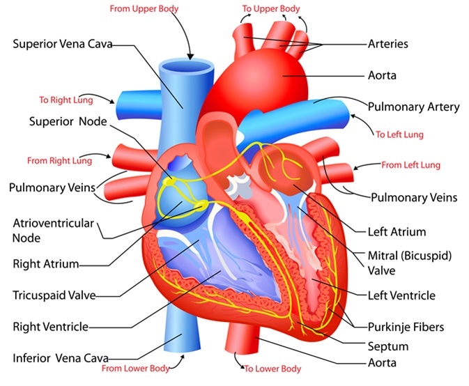
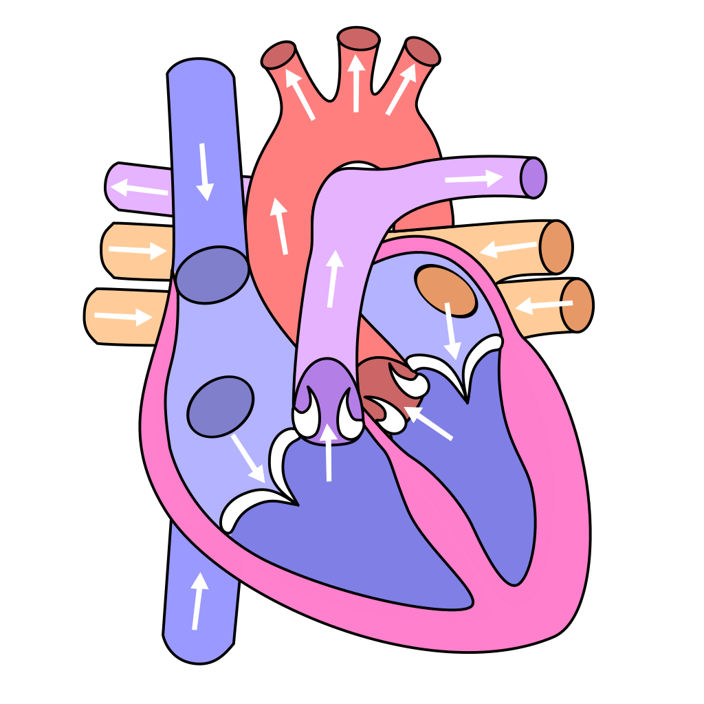
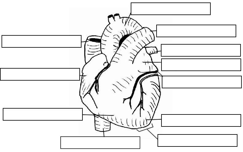
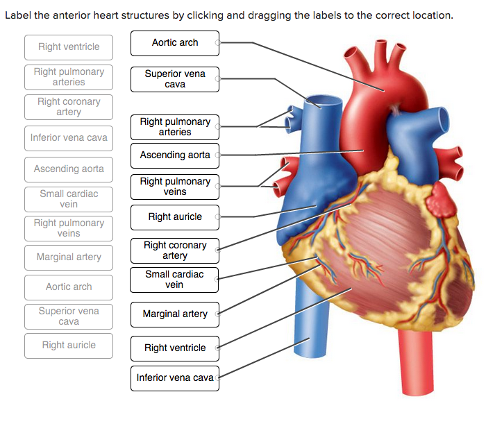
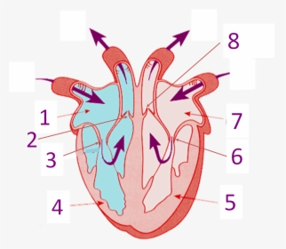
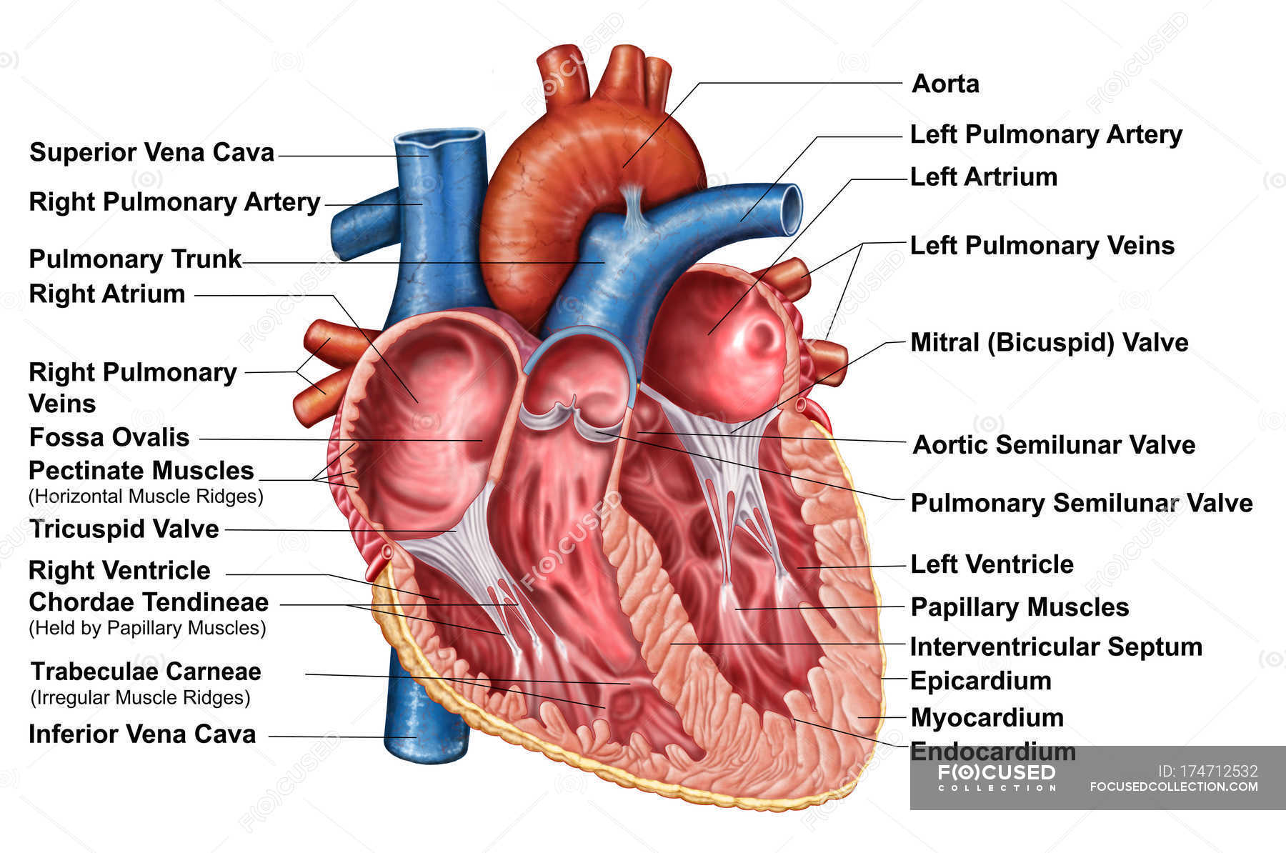
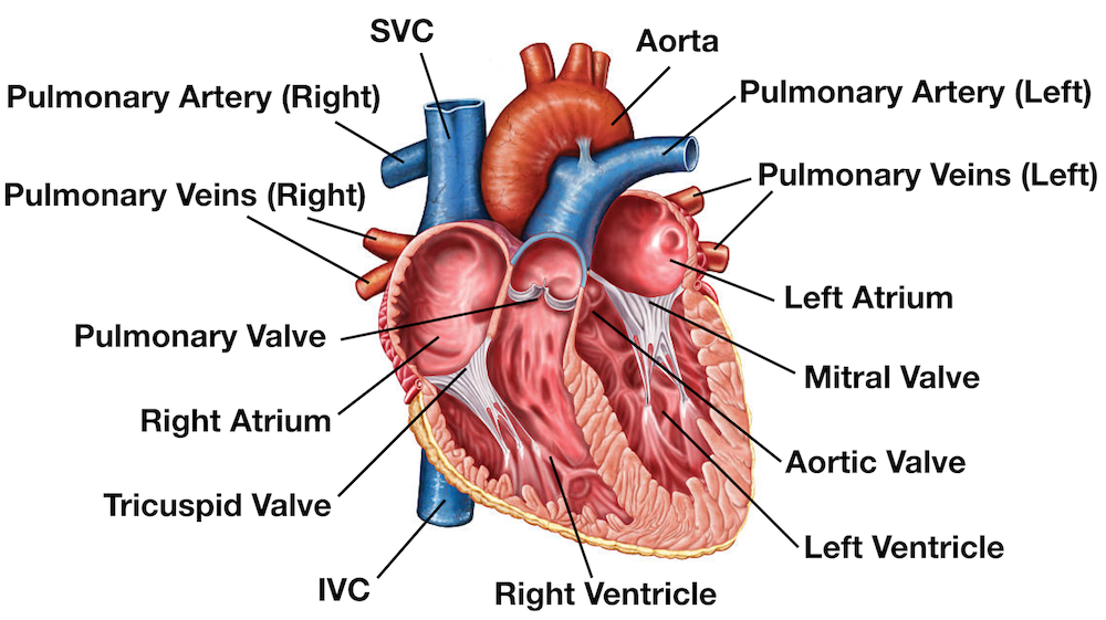
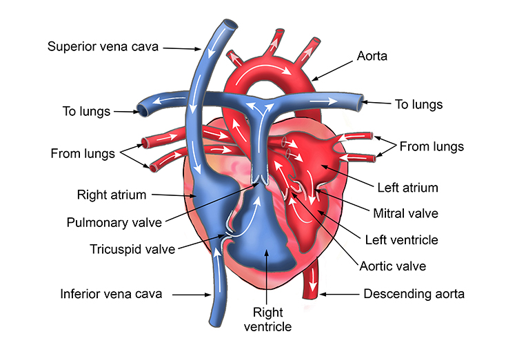
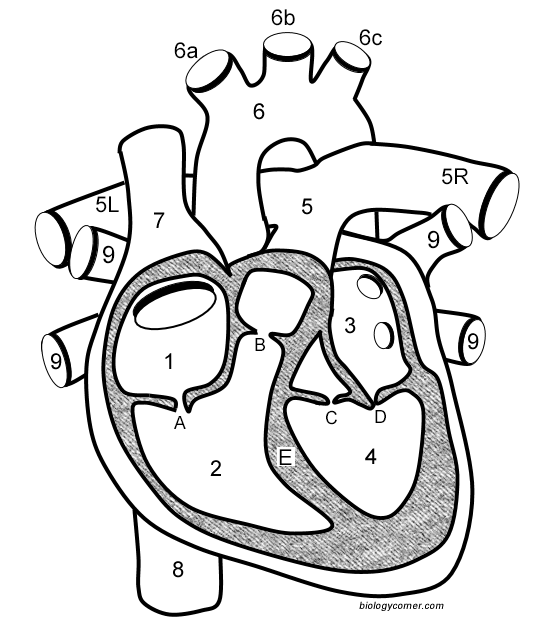
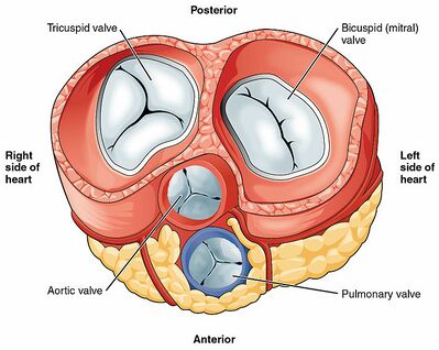
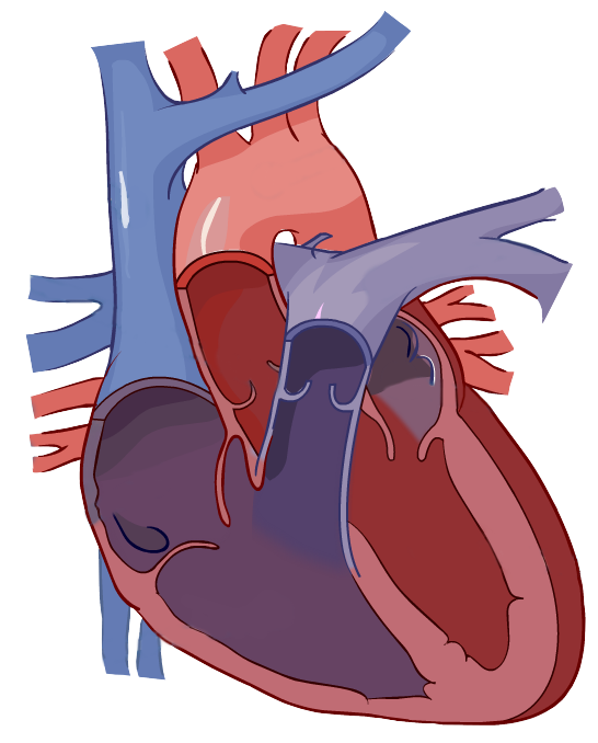
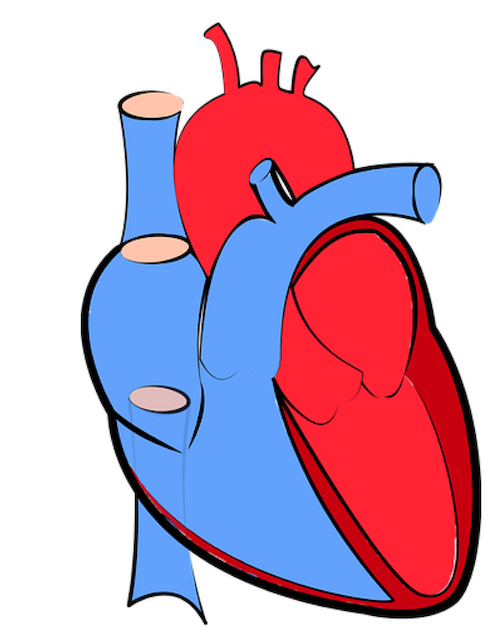
42 heart structure and labels
Heart: Anatomy and Function - Cleveland Clinic What are the parts of the heart's anatomy? The parts of your heart are like the parts of a house. Your heart has: Walls. Chambers (rooms). Valves (doors). Blood vessels (plumbing). Electrical conduction system (electricity). Heart walls Your heart walls are the muscles that contract (squeeze) and relax to send blood throughout your body. Heart: illustrated anatomy - e-Anatomy - IMAIOS Anatomical structures were labelled according to the actual Terminologia Anatomica. Anatomy of the human heart and coronaries: how to visualize anatomic structures This tool provides access to several medical illustrations, allowing the user to interactively discover heart anatomy.
Heart Anatomy: Labeled Diagram, Structures, Blood Flow ... 24 Feb 2022 — Function and anatomy of the heart made easy using labeled diagrams of cardiac structures and blood flow through the atria, ventricles, ...
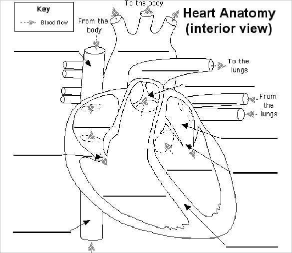
Heart structure and labels
Human Heart Diagram Labeled | Science Trends List Of Heart Structures Heart Chambers Ventricles - The bottom two heart chambers. Atra - The upper two heart chambers. Wall Of The Heart Sinoatrial Node - A collection of tissue that releases electrical impulses and defines the rate of contraction for the heart. Atrioventricular Bundle - The fibers which transmit cardiac impulses. Business News, Personal Finance and Money News - ABC News Oct 02, 2022 · Find the latest business news on Wall Street, jobs and the economy, the housing market, personal finance and money investments and much more on ABC News The Anatomy of the Heart, Its Structures, and Functions - ThoughtCo The heart is the organ that helps supply blood and oxygen to all parts of the body. It is divided by a partition (or septum) into two halves. The halves are, in turn, divided into four chambers. The heart is situated within the chest cavity and surrounded by a fluid-filled sac called the pericardium. This amazing muscle produces electrical ...
Heart structure and labels. Heart Diagram with Labels and Detailed Explanation - BYJUS Diagram of Heart. The human heart is the most crucial organ of the human body. It pumps blood from the heart to different parts of the body and back to the heart. The most common heart attack symptoms or warning signs are chest pain, breathlessness, nausea, sweating etc. The diagram of heart is beneficial for Class 10 and 12 and is frequently ... Structure and Function of the Heart - News-Medical.net Structure of the heart The heart wall is composed of three layers, including the outer epicardium (thin layer), middle myocardium (thick layer), and innermost endocardium (thin layer). The... Structure and function of the heart - BBC Bitesize The structure of the heart. If you clench your hand into a fist, this is approximately the same size as your heart. It is located in the middle of the chest and slightly towards the left. Heart Anatomy Labeling Game - PurposeGames.com This is an online quiz called Heart Anatomy Labeling Game There is a printable worksheet available for download here so you can take the quiz with pen and paper. Your Skills & Rank Total Points 0 Get started! Today's Rank -- 0 Today 's Points One of us! Game Points 19 You need to get 100% to score the 19 points available Actions
Heart Structure | BioNinja A heart is labelled as it would appear in a chest, so the left side of an image represents the right side of the heart (and vice versa). Structure of the Heart - SEER Training The human heart is a four-chambered muscular organ, shaped and sized roughly like a man's closed fist with two-thirds of the mass to the left of midline. The heart is enclosed in a pericardial sac that is lined with the parietal layers of a serous membrane. The visceral layer of the serous membrane forms the epicardium. Layers of the Heart Wall Methylene Blue - Structure, Formula, Preparation, Properties ... The various computed physical and chemical properties of methylene blue are that the molecular weight of methylene blue is 319.9, 0 hydrogen bond donor, 4 hydrogen bond acceptors, 1 rotatable bond, heavy atom count is 21, 0 formal charge, and 2 covalently bonded unit. SGLT2 inhibitor - Wikipedia The differences in the structures is relatively small. The general structure includes a glucose sugar with an aromatic group in the β-position at the anomeric carbon. In addition to the glucose sugar moiety and the β-isomeric aryl substituent the aryl group is composed of a diarylmethylene structure.
VP Online - Online Drawing Tool - Visual Paradigm VP Online is your all-in-one online drawing solution. Create professional flowcharts, UML diagrams, BPMN, ArchiMate, ER Diagrams, DFD, SWOT, Venn, org charts and mind map. Heart Information Center: Heart Anatomy Your heart has 4 chambers. The upper chambers are called the left and right atria, and the lower chambers are called the left and right ventricles. A wall of ... French degrees, LMD system and equivalences | Campus France For various "Grandes Ecoles" and business/engineering schools, the quality of training and diplomas may also be certified by independent organisations issuing accreditations or labels. Equivalences between French and foreign degrees Ch. 19 Circulatory System- heart Flashcards | Quizlet Place the labels in order denoting the flow of blood through the pulmonary circuit beginning with the right atrium and ending in the left atrioventricular valve. The first and last structures are given. Right atrium. 1. tricuspid valve. 2. right ventricle. 3. pulmonary valve. 4. pulmonary trunk. 5. pulmonary artery.
Metal Hammer | Louder Oct 04, 2022 · Wolfgang Van Halen suggests VH reunion is off the table, says former members are too "dysfunctional" to organise it anyway. By Liz Scarlett published 3 October 22 Exclusive Wolfgang Van Halen discusses the potential of a Van Halen reunion in Classic Rock magazine, and says playing VH songs at the Taylor Hawkins tribute concerts "delivered that catharsis" for him
heart | Structure, Function, Diagram, Anatomy, & Facts A thin layer of tissue, the pericardium, covers the outside, and another layer, the endocardium, lines the inside. The heart cavity is divided down the middle into a right and a left heart, which in turn are subdivided into two chambers. The upper chamber is called an atrium (or auricle), and the lower chamber is called a ventricle.
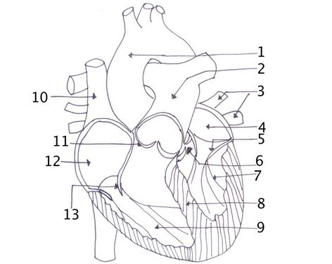
Lab 44- Heart Structure Flashcards | Quizlet Right side: 1.)Ligamentrum. 2.)left pulmonary artery. 3.)Pulmonary trunk. 4.)Left pulmonary veins. 5.)Auricle of left atrium. 6.)Grat cardiac vein. 7.)Anterior interventricular artery. Label the posterior heart structures by clicking and dragging the labels to the correct location.
Associate Members | Institute Of Infectious Disease and ... Associate membership to the IDM is for up-and-coming researchers fully committed to conducting their research in the IDM, who fulfil certain criteria, for 3-year terms, which are renewable.
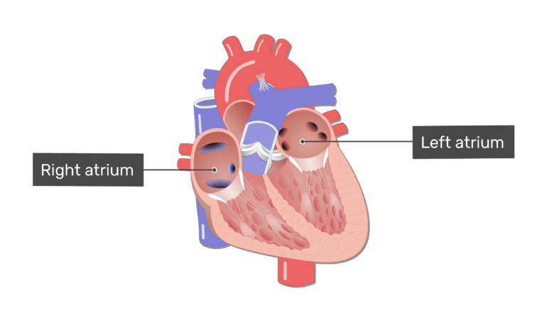
Heart anatomy: Structure, valves, coronary vessels | Kenhub Inside, the heart is divided into four heart chambers: two atria (right and left) and two ventricles (right and left).
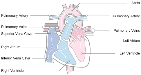
Label the heart - Science Learning Hub Label the heart Interactive Add to collection In this interactive, you can label parts of the human heart. Drag and drop the text labels onto the boxes next to the diagram. Selecting or hovering over a box will highlight each area in the diagram. pulmonary vein semilunar valve right ventricle right atrium vena cava left atrium pulmonary artery
Human Heart - Anatomy, Functions and Facts about Heart - BYJUS The human heart is divided into four chambers, namely two ventricles and two atria. The ventricles are the chambers that pump blood and atrium are the chambers that receive the blood. Among which, the right atrium and ventricle make up the "right portion of the heart", and the left atrium and ventricle make up the "left portion of the heart." 5.
Human Heart (Anatomy): Diagram, Function, Chambers, Location in ... - WebMD Chambers of the Heart The heart is a muscular organ about the size of a fist, located just behind and slightly left of the breastbone. The heart pumps blood through the network of arteries and...
Human Heart - Diagram and Anatomy of the Heart - Innerbody The heart contains 4 chambers: the right atrium, left atrium, right ventricle, and left ventricle. The atria are smaller than the ventricles and have thinner, less muscular walls than the ventricles. The atria act as receiving chambers for blood, so they are connected to the veins that carry blood to the heart.
147 Heart Anatomy With Labels Premium High Res Photos - Getty Images Browse 147 heart anatomy with labels stock photos and images available, or start a new search to explore more stock photos and images. of 3. NEXT.
Heart Labeling Quiz: How Much You Know About Heart Labeling? Here is a Heart labeling quiz for you. The human heart is a vital organ for every human. The more healthy your heart is, the longer the chances you have of surviving, so you better take care of it. Take the following quiz to know how much you know about your heart. Questions and Answers 1. What is #1? 2. What is #2? 3. What is #3? 4. What is #4?
Simple heart diagram labeled - Pinterest You can learn diagram of heart with labels and easy simple heart anatomy with heart structure. Learn to draw Simple heart diagram in very simple way and ...
Best way to draw and label the heart! | Heart Anatomy - YouTube In this lecture, Dr Mike shows the two best ways to draw and label the heart!
The Anatomy of the Heart, Its Structures, and Functions - ThoughtCo The heart is the organ that helps supply blood and oxygen to all parts of the body. It is divided by a partition (or septum) into two halves. The halves are, in turn, divided into four chambers. The heart is situated within the chest cavity and surrounded by a fluid-filled sac called the pericardium. This amazing muscle produces electrical ...
Business News, Personal Finance and Money News - ABC News Oct 02, 2022 · Find the latest business news on Wall Street, jobs and the economy, the housing market, personal finance and money investments and much more on ABC News
Human Heart Diagram Labeled | Science Trends List Of Heart Structures Heart Chambers Ventricles - The bottom two heart chambers. Atra - The upper two heart chambers. Wall Of The Heart Sinoatrial Node - A collection of tissue that releases electrical impulses and defines the rate of contraction for the heart. Atrioventricular Bundle - The fibers which transmit cardiac impulses.














Post a Comment for "42 heart structure and labels"