38 onion cells under microscope with labels
Onion cells hi-res stock photography and images - Alamy RF 2BN75YG - Under the microscope onion cells. RF MRGPTT - Onion epidermis under light microscope. Purple colored, large epidermal cells of an onion, Allium cepa, in a single layer. Photo. RM EBXPHH - Cell walls and organelles of onion bulb scale epidermis cells. RM CP074T - mitosis in a onion root, anaphasis. Plant Cell Under Microscope Observation : Grass cells under a ... Observe the slide under microscope. Observe the labeled diagram of plant cell. Draw the structure that you see under microscope!! Place the glass slide onto the stage. ... Observing Onion Cells under a Microscope - Blog, She Wrote from blogshewrote.org To look at a cell close up we need a microscope. Human cheek cell give blue and have dark ...
Onion Skin Cells - Investigation - Exploring Nature Observe the onion tissue under the microscope at 4x, 10x and 40x with lots of light (open diaphragm). Then slowly close the diaphragm while observing the image to find the best light for seeing cellular details. 6. Draw a section of onion skin cells at 10x magnification. Then switch to 40x and draw one cell and label it.

Onion cells under microscope with labels
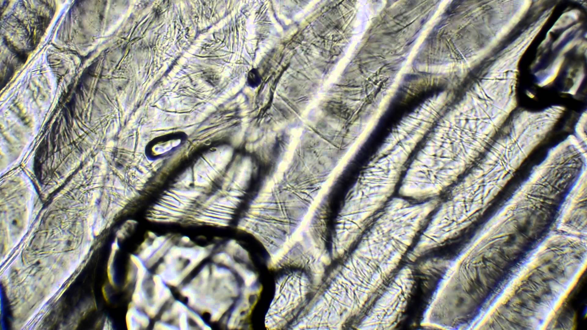
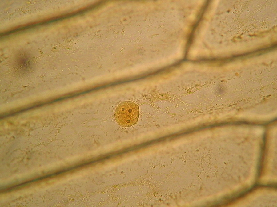
Onion Plant Cell Under Microscope Labeled - Ismael Dauila Studying cell tissues from an onion peel is a great exercise in using light microscopes and learning about plant cells, since onion cells are highly visible under a microscope, especially when stained correctly.onions are multicellular plant organisms, which basically means that they are made up of many cells. Onion Cells Microscope Stock Photos and Images - Alamy Onion skin cells under the microscope, horizontal field of view is about 0.61 mm ID: 2AM97C4 (RM) mitosis in a onion root, anaphasis ID: CP074T (RM) Red onion skin with oxalate crystals under the microscope, horizontal field of view is about 0.24mm ID: 2H9B7YK (RM) Onion epidermis (Allium cepa) showing cells and nucleus. Optical microscope X100. Onion Cell Microscope Slide Experiment - YouTube -- Materials: A Microscope, A Microscope Camera*, Microscope Slides, Coverslips, A Kitchen Knife, Pipettes*, Iodine Tincture, An Onion, Cutting Board -- I wrote a book about STEM! STEM Files is a...
Onion cells under microscope with labels. Onion Root Tip Mitosis - Stages, Experiment and Results - MicroscopeMaster · Using a blade or scalpel, carefully cut one or several roots from the onion and place them in a petri dish (or any other small clean container) · Prepare a water bath (about 55 degrees C) - The temperature should be maintained at 55 degrees C. This may be achieved by using a thermostatically controlled water bath Microscope Cell Lab: Cheek, Onion, Zebrina | SchoolWorkHelper The onion epidermis cell is the only cell that has a cell wall. In addition, it is the only cell that has a chloroplast, where photosynthesis can happen. The cheek epithelium cell is the only one that has centrioles, the barrel-shaped organelle that is responsible for helping organize chromosomes during cell division. Plant Cell Under Microscope Labeled 40X - Sadie Bermingham Microscopy and the interpretation of cell structures. (iv) describe how you applied the stain. Students will observe cheek cells under a microscope. Does anyone have a decent labelled diagram of a plant cell under an electron microscope? Onion cell) magnification (40x, 100x, or 400x) label all visible cell parts *use pencil or colored pencil. DOC The Onion Cell Lab - chsd.us Onion tissue provides excellent cells to study under the microscope. The main cell structures are easy to see when viewed with the microscope at medium power. For example, you will observe a large circular . nucleus. in each cell, which contains the genetic material for the cell. In each nucleus, are round bodies called . nucleoli
Lab: The Cell — The Biology Primer - Pinterest Observing onion cells under the microscope. For this microscope experiment, the thin membrane will be used to observe the cells. An easy beginner experiment. D Jessica Williams Ideas for Work Science For Kids Science And Nature Life Science Bee Facts Bee Boxes Backyard Beekeeping Bees And Wasps Bee Friendly Bugs And Insects Abejas D Daniel León DOC Plant and Animal Cells Microscope Lab - hillsboro.k12.oh.us Observe the onion cell under both low and high power. Make a drawing of one onion cell, labeling all of its parts as you observe them. (At minimum you should observe the nucleus, cell wall, and cytoplasm.) Cheek cells 1. To view cheek cells, gently scrape the inside lining of your cheek with a toothpick. DO NOT GOUGE THE INSIDE OF YOUR CHEEK! › lifestyleLifestyle | Daily Life | News | The Sydney Morning Herald The latest Lifestyle | Daily Life news, tips, opinion and advice from The Sydney Morning Herald covering life and relationships, beauty, fashion, health & wellbeing › bitesize › articlesCells and Reproduction - BBC Bitesize Onion cells are easy to see using a light microscope. ... A small tube placed under the skin of the upper arm. ... Five small tubes with labels and stoppers or lids Cress seeds Labels Cotton wool ...
› your-liverYour liver is essential to your life. The Canadian Liver ... This is a myth. Jaundice can be an early warning sign of liver disease. Many babies have “newborn jaundice” lasting three to five days after birth because their liver is not yet fully developed, however, jaundice that does not clear up after 14 days of life, dark urine and/or pale stools, an enlarged abdomen and vomiting are signs that your baby should be seen by his or her doctor. What Does a Worm Look Like Under a Microscope? The Earthworm Under The Microscope With an electron microscope, you'll be able to see both the outside skin and internal (through dissection) anatomy of the earthworm. You will need the following for this experiment: Microscope Earthworm specimen Glass slide A pair of tweezer Water Alcohol solution Procedure: Onion Cells Under a Microscope - Requirements/Preparation/Observation Add a drop of iodine solution on the onion membrane (or methylene blue) Gently lay a microscopic cover slip on the membrane and press it down gently using a needle to remove air bubbles. Touch a blotting paper on one side of the slide to drain excess iodine/water solution, Place the slide on the microscope stage under low power to observe. Onion Peels Observed Under the Microscope | Confirmation Point Onion Peels Observed Under the Microscope Cells present in onion peel can be observed under microscope. For this onion peels are first isolated. For this experiment outer most scale of the onion is removed and is cut into four equal halves. It is a monocot plant. Then with the help of a pairs of forcep the scale of onion is peeled out.
What organelles are in an onion cell? - Biology Stack Exchange You cannot see most of these as they appear translucent as well as being too small to see under the light microscope. You need an electron microscope to view these. Note: chloroplasts are not present in an onion cell as it is not a photosynthesising cell. This is a typical onion cell slide with labels: Share Improve this answer
Onion Epidermis - kuensting.org Onion epidermal cells, iodine stain, 400X. The nucleus of an onion epidermal cell, 1000X magnification. ...
Blood under microscope labeled Search: Leaf Cell Under Microscope Labeled. Animal cells do not have a cell wall nucleus (if Label the square "Haploids" The leaf is the site of two major processes: gas exchange and light capture, which lead to photosynthesis Biology 130 Lab 3 Light Microscope Images Lab manual exercise 1 lab manual exercise 1 virtual biology labs study botany ...
en.wikipedia.org › wiki › CeleryCelery - Wikipedia A celery stalk readily separates into "strings" which are bundles of angular collenchyma cells exterior to the vascular bundles. Wild celery, Apium graveolens var. graveolens, grows to 1 m (3 ft 3 in) tall. Celery is a biennial plant that occurs around the globe. It produces flowers and seeds only during its second year.
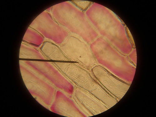
Observing Onion Cells Under The Microscope » Microscope Club The main onion cell structures are quite easy to observe under medium magnification levels when using a light microscope. The cells look elongated, similar in appearance- color, size, and shape- have thick cell walls, and a nucleus that is large and circular in shape. How to observe onion cells under a microscope
Onion Cell Under Microscope Labeled a scientist is observing onion cells and human cheek cells under a microscope label the cell wall, nucleus, and cytoplasm draw and label your observations: label the nucleus, cell wall, and cytoplasm of one onion cell students know the characteristics that distinguish plant cells from animal cells, including chloroplasts and cell walls once you …
Onion Cell Diagram Labeled Pdf [PDF] - thesource2.metro Set your multimeter to measure current in the 20 mA range (the dial setting labeled "20m" on the right). Plug the multimeter's black probe into the port labeled COM. Plug the multimeter's red probe into the port labeled VΩmA. Use a red alligator clip lead to connect the multimeter's red probe to the positive (+) terminal of the 9 V battery.
Hypothesis onion cell will be visible when viewed in - Course Hero 12.Again, looking from the SIDE of the microscope, rotate the lenses to the highest-powered lens (40x objective). If you need to, use the fine focus knob (the smallest knob) to get the image into focus. You should see a dark blob in the middle of each cell. 13.In your lab notebook, draw a picture of what you see. Label the picture "Onion skin cells 400x".
Onion Cell Lab Report.docx - Onion Cell Lab Report By station, remove the single layer of epidermal cells from inner side of the scale leaf. 3(Place the single layer of onion on a glass slide. 4(Place a drop of iodine stain on your onion tissue. 5(Put the cover slip on the stained tissue and gently tap out any air bubbles. 6(Observe the cells under the microscope and see you results.
Microscopes and Cells - Biology I: Introduction to Cell and Molecular ... View the leaf under low, medium, and high power objectives, and then draw the cells in Figure 2.2, along with any organelles you can see. Be sure to label the chloroplasts, the cell membrane, and the cell wall. Onion Epithelial Cells. Use half of a slide to examine onion cells. Cut a small piece of onion and break it by bending it in half.
› health › alopecia-areataAlopecia Areata: Causes, Symptoms, and Diagnosis - Healthline Mar 01, 2022 · Alopecia areata is an autoimmune condition.An autoimmune condition develops when the immune system mistakes healthy cells for foreign substances. Normally, the immune system defends your body ...
Onion Cells Under a Microscope (100x-2500x) - YouTube 567 views Feb 24, 2021 In this video you will see onion cells under a microscope (100x-2500x) as is, without any coloring. To observe the onion cells the thin membrane is used. It can easil ...more...
› 44090147 › Cambridge(PDF) Cambridge International AS and A Level Biology ... BIO1: Maintaining a Balance 1. Most organisms are active in a limited temperature range IDENTIFY THE ROLE OF ENZYMES IN METABOLISM, DESCRIBE THEIR CHEMICAL COMPOSITION AND USE A SIMPLE MODEL TO DESCRIBE THEIR SPECIFICITY ON SUBSTRATES
assist.asta.edu.auASSIST | Australian school science information support for ... Australian School Science Information and Support For Teachers and Technicians
NCERT Class 9 Science Lab Manual - Slide of Onion Peel and Cheek Cells ... The cells observed under the microscope do not have cell wall and big vacuoles, these are the cells of animal. Precautions. Use unused/new toothpick for scraping of cheek cells. Placing of coverslip should be done carefully to avoid air bubbles. Avoid overstaining. Use clean/dry mounted slide while placing it under the lens of the microscope.
Onion Cell Microscope Slide Experiment - YouTube -- Materials: A Microscope, A Microscope Camera*, Microscope Slides, Coverslips, A Kitchen Knife, Pipettes*, Iodine Tincture, An Onion, Cutting Board -- I wrote a book about STEM! STEM Files is a...


Post a Comment for "38 onion cells under microscope with labels"