40 diagram of a human cell with labels
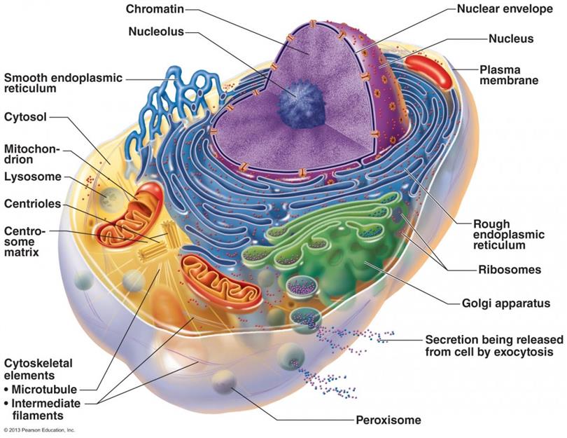
PDF Human Cell Diagram, Parts, Pictures, Structure and Functions Diagram of the human cell illustrating the different parts of the cell. Cell Membrane The cell membraneis the outer coating of the cell and contains the cytoplasm, substances within it and the organelle. It is a double-layered membrane composed of proteins and lipids. Cell: Structure and Functions (With Diagram) - Biology Discussion Eukaryotic Cells: 1. Eukaryotes are sophisticated cells with a well defined nucleus and cell organelles. 2. The cells are comparatively larger in size (10-100 μm). 3. Unicellular to multicellular in nature and evolved ~1 billion years ago. 4. The cell membrane is semipermeable and flexible. 5. These cells reproduce both asexually and sexually.
Label Diagram Human Body Illustrations & Vectors - Dreamstime Download 201 Label Diagram Human Body Stock Illustrations, Vectors & Clipart for FREE or amazingly low rates! New users enjoy 60% OFF. 190,035,120 stock photos online. ... Animal cell structure anatomy infographic diagram. With parts flat vector illustration design for biology science education school book concept microbiology.
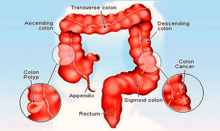
Diagram of a human cell with labels
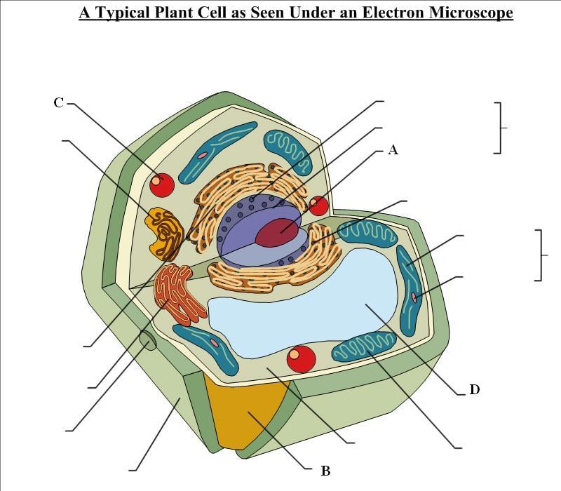
Learn the parts of a cell with diagrams and cell quizzes For this exercise we'll start with an image of a cell diagram ready labeled. Study this and make sure that you're clear about which structure is found where. Cell diagram unlabeled It's time to label the cell yourself! As you fill in the cell structure worksheet, remember the functions of each part of the cell that you learned in the video. Labeled Plant Cell With Diagrams | Science Trends Plant cells contain many organelles such as ribosomes, the nucleus, the plasma membrane, the cell wall, mitochondria, and chloroplasts. In addition, plant cells differ from animal cells in a number of key ways. Examining a diagram of the plant cell will help make the differences clearer. Let's go over the individual components of plant cells ... Cells Diagram | Science Illustration Solutions - Edrawsoft Cells Diagram Symbols Edraw software offers you lots of symbols used in cells diagram like cell structure, paramecium, squamous cell, cell division, bacteria, cell membrane, eggs, sperm, zygote, an animal cell, SARS, tobacco mosaic, adenovirus, coliphage, herpesvirus, AIDS, pollen, plant cell model, onion tissue, etc. Cells Diagram Examples
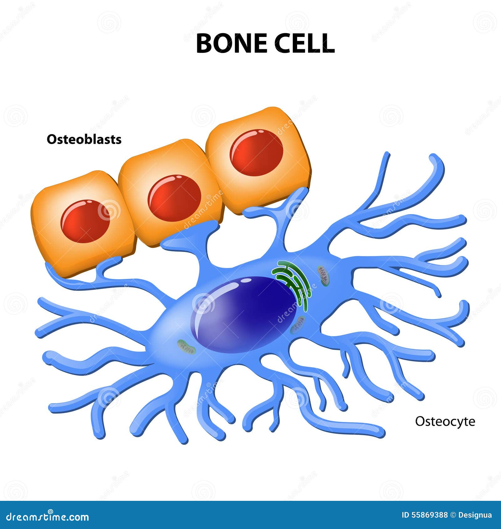
Diagram of a human cell with labels. Plant Cell Diagram | Science Trends A plant cell diagram, like the one above, shows each part of the plant cell including the chloroplast, cell wall, plasma membrane, nucleus, mitochondria, ribosomes, etc.A plant cell diagram is a great way to learn the different components of the cell for your upcoming exam. Plants are able to do something animals can't: photosynthesize.Plant cells are able to do this because plant cells have ... Labelled Diagram Of A Human Cell Bone Cell Labeled Diagram Animal Cell ... Labelled Diagram Of A Human Cell Bone Cell Labeled Diagram Animal Cell Free Printable To Label Find this Pin and more on Biology by Guinahi Douhe. Cell Structure Structure And Function Plasma Membrane Classroom Management Tips Label Image Animal Cell Application Letters Plant Cell Cell Wall More information ... More information Animal Cells: Labelled Diagram, Definitions, and Structure - Research Tweet Animal Cells Organelles and Functions. A double layer that supports and protects the cell. Allows materials in and out. The control center of the cell. Nucleus contains majority of cell's the DNA. Popularly known as the "Powerhouse". Breaks down food to produce energy in the form of ATP. Human Cell Organelles Labeling Diagram | Quizlet Human Cell Organelles Labeling STUDY Learn Flashcards Write Spell Test PLAY Match Gravity Created by Mackenna_Rios5 Terms in this set (8) Vesicles Transports molecules between organelles and the cell membrane Ribosome Makes Protein Mitochondria Makes ATP Smooth ER Makes lipids and vesicles Lysosomes
human hand diagram with labels bones diagram labeled hand anatomy skeleton left human list body chart labels diagrams worksheets worksheeto worksheet via buzzle. IB Biology Notes - 2.3 Eukaryotic Cells ibguides.com. biology cell eukaryotic animal cells ib notes liver diagram drawing draw biological ultrastructure label example plant drawings annotated membrane monday. What ... animal bone cell diagram labeled blood microscope under smear peripheral cells labeled histology seen shutterstock illustration lab. Christian Revolution Animal Cell Diagram With Labels And Functions . cell animal diagram labels structure project cells. Connective Tissue - BIOLOGY4ISC biology4isc.weebly.com diagram of a cell labeled Cell cells science inside discover constitution animal structure human anatomy biology body kidsdiscover diagram project peek issues function plant parts Quia - Meiosis Illustration Identification. 18 Pictures about Quia - Meiosis Illustration Identification : A typical cell, labeled diagram. | Alila Medical Images, CELL - Labelled diagram and ... Labeled Diagram of the Human Kidney - Bodytomy Labeled Diagram of the Human Kidney The human kidneys house millions of tiny filtration units called nephrons, which enable our body to retain the vital nutrients, and excrete the unwanted or excess molecules as well as metabolic wastes from the body. In addition, they also play an important role in maintaining the water balance of our body.
Labeled Diagrams of the Human Brain You'll Want to Copy Now All the functions are carried out without a single glitch and before you even bat an eyelid. The following are the different regions of the human brain and their functions. Labeled Diagrams of the Human Brain Central Core The central core consists of the thalamus, pons, cerebellum, reticular formation and medulla. Human Cells Printables and Diagrams - The Successful Homeschool These cells include: leukocytes, haematids, thrombocytes, ovum, sperm, sarcomeres, enterocytes, neurons, osteocytes, hepatocytes. They will learn the parts of a cell thanks to a labeled diagram. They will get to see what blood looks like under a microscope without needing to own a microscope. They get to color a cell and then label the parts. Human Cell Diagram, Parts, Pictures, Structure and Functions Diagram of the human cell illustrating the different parts of the cell. Cell Membrane. The cell membrane is the outer coating of the cell and contains the cytoplasm, substances within it and the organelle. It is a double-layered membrane composed of proteins and lipids. The lipid molecules on the outer and inner part (lipid bilayer) allow it to ... label diagram of plant cell Nucleus cell biology exams. label diagram of plant cell. animal cell to label we have 9 Pics about animal cell to label like Nucleus, the commanding centre of the cell ~ Biology Exams 4 U, 12 Best Images of Human Brain Diagram Worksheet - Human Brain Anatomy and also Interactive Eukaryotic Cell Model. Here it is:
detail colon cancer | Anatomy System - Human Body Anatomy diagram and chart images
Circulatory System Labeled Diagram stock illustrations The urinary system. The human urinary system medical illustration with internal organs. Double circulation vector illustration. Labeled educational... Double circulation vector illustration. Labeled educational blood route scheme. Lung capillaries, pulmonary artery, aorta, vein and vena cava diagram.
Cell, Diagram of | ClipArt ETC
File:Diagram human cell nucleus tr.svg - Wikimedia Commons Description. Diagram human cell nucleus tr.svg. en: A diagram of a human cell nucleus, with Turkish labels. Translated version of File:Diagram human cell nucleus.svg, originally created and all rights released by Mariana Ruiz ( User:LadyofHats ). This image is also released to the public domain. az: İnsan hüceyrə nüvəsinin sxematik rəsmi ...

The human egg cell explained for egg donors | Altrui
Human cell diagram | Human cell diagram, Cell diagram, Human cell structure Human Cell Diagram Human Cell Model. Made from a painted foam ball, and clay. • •Shelby James• Projects Science Cells Science Biology Life Science Biology Lessons Study Biology Teaching Biology Human Cell Structure A basic, living animal cell with it's organselles (mini organs) all working together to keep it functioning at it's peak.

Learn all about plant cells with this printable illustration. in 2020 | Plant cell, Plant and ...
A Labeled Diagram of the Animal Cell and its Organelles A Labeled Diagram of the Animal Cell and its Organelles There are two types of cells - Prokaryotic and Eucaryotic. Eukaryotic cells are larger, more complex, and have evolved more recently than prokaryotes. Where, prokaryotes are just bacteria and archaea, eukaryotes are literally everything else.

September 2010 ~ Pass. Science. Solutions.
Blood Cell Diagram Pictures, Images and Stock Photos Realistic vector 3d picture of blood cells in human vein Blood Cells Poster Blood cells types. Editable vector illustration isoated on white background. Erythrocytes, plateletes, leukocytes, lymphocytes, monocytes and more. Educational medical poster in landscape format. blood cell diagram stock illustrations Blood Cells Poster Blood cells types.

Cell Review Guide Answers | Human cell diagram, Human cell structure, Animal cell parts
Liver Diagram with Detailed Illustrations and Clear Labels - BYJUS Liver Diagram with Detailed Illustrations and Clear Labels Biology Important Diagrams Liver Diagram Liver Diagram The liver is one of the most important organs in the human body. Anatomically, the liver is a meaty organ that consists of two large sections called the right and the left lobe.

Explain the nucleus of a cell with a neat labeled diagram - Science - Cell - Structure and ...
03 Label the Cell Diagram | Quizlet Start studying 03 Label the Cell. Learn vocabulary, terms, and more with flashcards, games, and other study tools.

Follow me: Arts
label diagram of tissue cells label diagram of tissue cells Basic Histology -- Smooth Muscle, Longitudinal Section we have 9 Pics about Basic Histology -- Smooth Muscle, Longitudinal Section like Pseudostratified Ciliated Columnar Epithelium Shows Cilia Ciliated, Human Anatomy Lab Exercises Tissues Recognition and Function Flashcards and also Nervous Tissue - YouTube.

Diagram shows one of the cells in the human body. | TeacherNotes4U
Cell Organelles- Definition, Structure, Functions, Diagram - Microbe Notes A cell wall is multilayered with a middle lamina, a primary cell wall, and a secondary cell wall. The middle lamina contains polysaccharides that provide adhesion and allow binding of the cells to one another. After the middle lamina is the primary cell wall which is composed of cellulose.
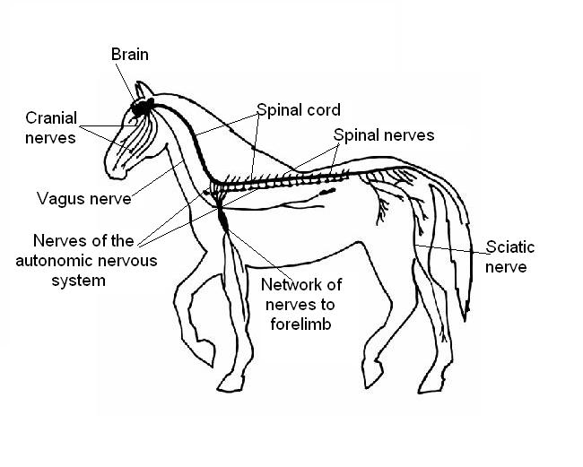
The Anatomy and Physiology of Animals/Nervous System Worksheet/Worksheet Answers - WikiEducator
cell diagram unlabeled Cell Diagram Unlabeled - Diagram Media diagramedia.blogspot.com. 2.2.1 Draw And Label A Diagram Of The Ultrastructure Of EColi As An . diagram label draw ecoli ultrastructure prokaryote example. Nervous system review 9-1 to 9.10. Label the model human cell. Human diagram skeleton printable labeled anatomy blank labelled unlabeled ...

Questions And Answers On Labeled/Unlebled Diagrams Of A Human Cell : Animal Cell Diagram ...
Cells Diagram | Science Illustration Solutions - Edrawsoft Cells Diagram Symbols Edraw software offers you lots of symbols used in cells diagram like cell structure, paramecium, squamous cell, cell division, bacteria, cell membrane, eggs, sperm, zygote, an animal cell, SARS, tobacco mosaic, adenovirus, coliphage, herpesvirus, AIDS, pollen, plant cell model, onion tissue, etc. Cells Diagram Examples
25 Human Cell Diagram To Label - Wiring Database 2020
Labeled Plant Cell With Diagrams | Science Trends Plant cells contain many organelles such as ribosomes, the nucleus, the plasma membrane, the cell wall, mitochondria, and chloroplasts. In addition, plant cells differ from animal cells in a number of key ways. Examining a diagram of the plant cell will help make the differences clearer. Let's go over the individual components of plant cells ...
Biological Quiz On Cell Parts And Functions - ProProfs Quiz
Learn the parts of a cell with diagrams and cell quizzes For this exercise we'll start with an image of a cell diagram ready labeled. Study this and make sure that you're clear about which structure is found where. Cell diagram unlabeled It's time to label the cell yourself! As you fill in the cell structure worksheet, remember the functions of each part of the cell that you learned in the video.
Osteoblast Cartoons, Illustrations & Vector Stock Images - 23 Pictures to download from ...

Pin by Nicole Vardiabasis on Biology | Human cell diagram, Human cell structure, Animal cell parts
Animal Cell Features
Post a Comment for "40 diagram of a human cell with labels"