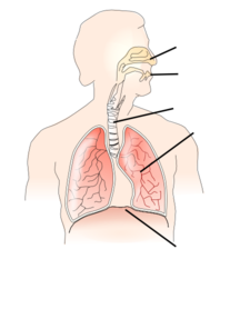
42 lungs pictures with labels
How the Lungs Work - The Lungs | NHLBI, NIH Español Your lungs are the pair of spongy, pinkish-gray organs in your chest. When you inhale (breathe in), air enters your lungs, and oxygen from that air moves to your blood. At the same time, carbon dioxide, a waste gas, moves from your blood to the lungs and is exhaled (breathed out). This process, called gas exchange, is essential to life. Creating Model Working Lungs: Just Breathe - Activity A model of the lungs. Pull the diaphragm (balloon) down (that is, away from the lungs) in order to inflate the lungs. (Note: This makes the chest cavity larger and decreases the pressure.) Push the diaphragm (balloon) in (towards the lungs) in order to deflate the lungs.
[Unlabeled Diagram Of The Lungs] - 16 images - the anatomy and ... [Unlabeled Diagram Of The Lungs] - 16 images - class blog july 2012, what purpose do heart valves serve quora, what are the most common causes of phlegm in the lungs, understanding the anatomy of the respiratory system a d a m ondemand,
Lungs pictures with labels
Chest anatomy illustrations - e-Anatomy - IMAIOS Anatomy of the chest and the lungs: anatomical illustrations. This e-Anatomy module presents an illustrated anatomy of the lungs, trachea, bronchi, pleural cavity and pulmonary vessels. This thoracic and pulmonary anatomy tool is especially designed for students of anatomy (medical and paramedical studies). › Pure-Encapsulations-Calcium-DAmazon.com: Pure Encapsulations Calcium-D-Glucarate ... About this item . Detoxification: Calcium-D-Glucarate, a patented form of glucaric acid, is a nutrient with the potential to support healthy detoxification, lipid metabolism, and cellular function.* Body Cavities and Membranes: Labeled Diagram, Definitions The ventral cavity is highlighted in red and labeled with a star below. You can see how the ventral cavity is situated in front of or anterior to the dorsal cavity. The ventral cavity houses the contents within the chest, abdomen, and pelvis.
Lungs pictures with labels. What is Lung Fibrosis? (with pictures) - Info Bloom Lung fibrosis, also known as pulmonary fibrosis, is a serious medical condition that involves scarring of the lung tissue. This condition occurs when the alveoli, or air sacs, become inflamed and develop scars on the lung tissue in an attempt to repair themselves. There is no known cure or way to reverse the scarring on the lungs, so treatment ... › Use-Voldyne-5000How to Use Voldyne 5000 (with Pictures) - wikiHow Jun 25, 2021 · Hold the Voldyne 5000 in an upright position. Keep the device in an upright position with all labels facing you. Keep the device as level as possible so that the procedure will work correctly and all readings will be accurate. › photos › male-human-anatomyMale Human Anatomy Diagram Pictures, Images and Stock Photos Human anatomy, back injury or disease, medical concepts. Three main curvatures of the spine disorders or deformities on male body: lordosis, kyphosis and scoliosis 3D rendering illustration. Human anatomy, back injury or disease, medical concepts. male human anatomy diagram stock pictures, royalty-free photos & images Labeled imaging anatomy cases | Radiology Reference Article ... URL of Article. This article lists a series of labeled imaging anatomy cases by body region and modality. On this page: Article: Brain. Head and neck. Spine. Chest. Abdomen and pelvis.
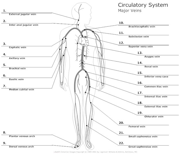
Heart: illustrated anatomy - e-Anatomy - IMAIOS This interactive atlas of human heart anatomy is based on medical illustrations and cadaver photography. The user can show or hide the anatomical labels which provide a useful tool to create illustrations perfectly adapted for teaching. Anatomy of the heart: anatomical illustrations and structures, 3D model and photographs of dissection. Free Heart Worksheets for Human Anatomy Lessons Print out sheet of the human heart with labels - This fun heart worksheet shows kids the different parts of the heart. They'll learn about the left ventricle, the left atrium, the tricuspid valve, and more. Human Heart Clipart - There is a coloring page, heart labeling worksheet and heart anatomy chart. What is the Bronchial Tree? (with pictures) - Info Bloom The bronchial tree begins with the primary bronchi and ends with the alveoli. The bronchial tree is an essential part of the respiratory system. It consists of several interacting structures, such as the bronchi, bronchioles, and alveoli. These structures work together to provide a network system between the lungs and the trachea. Picture Of Circulatory System With Labels / 11 00 Select from 1315 premium circulatory system label of . The heart (cardiovascular), lungs (pulmonary), and arteries . Huge collection, amazing choice, 100+ million high quality, affordable rf and rm images. You'll love the human heart circulatory system diagram chart medical. Find the perfect circulatory system stock photo.
Graphic warning labels on cigarettes could have prevented hundreds of ... adding warning labels like these with graphic depictions of the negative health consequences of cigarette smoking could have averted thousands of smoking-related deaths if approved as originally... Circulatory System Anatomy, Diagram, & Function - Healthline The circulatory system works thanks to constant pressure from the heart and valves throughout the body. This pressure ensures that veins carry blood to the heart and arteries transport it away ... Respiratory system quizzes and labeled diagrams | Kenhub Take a look at the labeled diagram of the respiratory system above. As you can see, there are several structures to learn. Spend a few minutes reviewing the name and location of each one, then try testing your knowledge by filling in your own diagram of the respiratory system (unlabeled) using the PDF download below. Respiratory system unlabeled WHMIS 2015 - Pictograms : OSH Answers Most pictograms have a distinctive red "square set on one of its points" border. Inside this border is a symbol that represents the potential hazard (e.g., fire, health hazard, corrosive, etc.). Together, the symbol and the border are referred to as a pictogram. Pictograms are assigned to specific hazard classes or categories.
Hilum of the Lung: Definition, Anatomy, and Masses A test called a mediastinoscopy (a surgical procedure in which a surgeon is able to explore the area between the lungs, including the hilar lymph nodes) may be needed to better visualize the region or to obtain a biopsy sample, though PET scanning has replaced the need for this procedure in many cases. 4 Hilar Enlargement/Hilar Masses
Diagram of Human Heart and Blood Circulation in It Ventricle contracts and pushes the blood into the pulmonary artery that sends blood to your lungs from where oxygen-rich blood returns to the left ventricle and the process continues. Exterior of the Human Heart A heart diagram labeled will provide plenty of information about the structure of your heart, including the wall of your heart.
Free Respiratory System Worksheets and Printables Respiratory System Doodle Labeled Coloring Page - This coloring page includes wonderful details about the respiratory system such as an explanation about how the diaphragm contracts and a close-up image of the lung alveoli. If your kids love to color, this is the perfect worksheet for you! Respiratory System Notebooking Pages
Pictures of Tips for Living With Pulmonary Arterial Hypertension Keep your go-to kitchen items on the countertop. Put laundry soap next to or just above the machine. Move your bathroom products from under the sink up to the medicine cabinet. You want your chest ...
This Is What COPD Looks Like in the Lungs - Verywell Mind Lungs are divided into lobes, balloon-like structures that receive air from the bronchial tubes. The left lung (pictured) has two lobes and the right lung has three. A normal human lung is pink and spongy, filled with an intricate system of airways and thousands of tiny alveoli sacs. 4 Picture of Human Right Lung With Emphysema
Histology, Lung - StatPearls - NCBI Bookshelf The lung is identified, dissected en-block, weighed, and labeled. Later the lung is perfused with 10% formalin through the trachea to the physiological peak inspiration level. Underinflation or over inflation should be prevented. This helps in proper assessment without any artifacts and over/under the judgment of the tissue structure.
› photos › human-throat-anatomyHuman Throat Anatomy Pictures, Images and Stock Photos Lungs and human body Stylized male human body with lungs human throat anatomy stock illustrations Lungs and human body Throat diseases, chromolitograph, published in 1897 Throat diseases: 1) Membranous tan of the larynx and trachea (cross-section view from behind); 2) Tuberculous laryngeal phthisis (cross-section view from behind); 3) Laryngeal ...
Anatomy of The Human Ribs - With Full Gallery Pictures! The Anatomy of the Human Ribs (costae) are one of the integral parts of the chest wall; they make up the lateral part of our body, its anterior and posterior wall and they entirely build the lateral parts of the chest wall. The anatomy of the human ribs is made up of 24 ribs. These ribs are parted in 12 pairs (each on the left and right side of ...
Lung - Pathology Outlines - Histology Positive stains. Mucous cell (goblet cell): MUC2 and MUC5AC. Basal cell: cytokeratin 5/6, cytokeratin 34 beta E12, p63 and p40. Neuroendocrine cell (Kulchitsky cell): CD56, synaptophysin and chromogranin A ( Mills: Histology for Pathologists, 5th Edition, 2019 ) Alveolar epithelium: cytokeratin 7.
› watchFive Senses Song | Song for Kids | The Kiboomers - YouTube The Kiboomers! Five Senses! Kids Songs for Preschool!★Get this song on iTunes: ...
Idiopathic Pulmonary Arterial Hypertension - Drugs.com A V/Q scan is a two-part test which takes pictures of your lungs to look for certain lung problems. During the perfusion part of the test, radioactive dye is put into your vein (blood vessel). The blood carries the dye to the blood vessels in your lungs. Pictures are taken to see how blood flows in your lungs.
› health › paint-fumesImpact of Paint Fumes on Your Health & How to Minimize Exposure Jul 08, 2019 · When shopping for paint, check the labels to get an idea of a product’s VOC levels. ... Poison Oak Rash: Pictures and Remedies. Medically reviewed by Chris Young, DNP, RN, NE-BC, NPD.
Bone markings [the complete list] | Kenhub The body, or diaphysis (dia- meaning "through" or "throughout") refers to the central shaft running between the proximal and distal ends of the bone. The articular surface (can be more than one) is the area of the bone that comes in close proximity with the neighbouring bones.
› smokers-lungs-vs-normalSmoker's Lungs vs. Normal Healthy Lungs - verywellmind.com Feb 09, 2022 · The healthy lungs of someone who doesn't smoke appear pink or red in color, whereas the lungs of someone who smokes can turn black or gray over time, depending on how heavily they smoke. The cause of the discoloration is the black-pigmented tar (particulate matter that is created when you burn tobacco) that is inhaled when you puff a cigarette.
Your Digestive System in Pictures - Verywell Health StA-gur Karlsson/E+/Getty Images. You will find that you may be able to ease some of the anxiety that goes along with not feeling well by having a good understanding of what your digestive system looks like inside of you. Looking at pictures of your GI tract can help you to pinpoint where symptoms such as abdominal pain may be coming from.
Normal chest imaging examples | Radiology Reference Article ... HRCT chest. example 1 : non-contrast, supine, prone and expiratory. CT pulmonary angiogram (CTPA) example 1 : excellent pulmonary arterial / venous differentiation. example 2: average pulmonary arterial / venous differentiation. example 3: spectral CTPA. CT coronary arteries (CTCA) right dominant: example 1, example 2.


Post a Comment for "42 lungs pictures with labels"